

We often think of skincare in terms of surface-level fixes. Creams that hydrate the top layer or serums that temporarily tighten. However, dermatologists and regenerative medicine doctors will tell you the real transformation occurs at a deeper biological level. The next generation of skincare treatments isn’t simply covering up damage; new techniques support the body in rebuilding itself, just as it did when we were children. It’s why so many of the world’s top skin health experts are so excited about Muse cells for skin regeneration.
Muse cells thrive in the harsh environments of inflammation and injury. They are the body’s natural emergency responders, and they are changing the conversation around how we treat wounds, scarring, and the inevitable aesthetic impact of aging. No longer is the conversation simply centered on temporary improvements. Patients across the U.S. are turning to Muse cells for lasting skin health.
What Are Muse Cells?
To understand why Muse cells are making waves in dermatology, we must first explain their unique characteristics. Muse cells are a distinct subpopulation of mesenchymal stem cells (MSCs) that were discovered by Professor Mari Dezawa at Tohoku University in Japan in 2010.
While most cells are specialized, a skin cell is a skin cell, a muscle cell is a muscle cell, Muse cells are naturally occurring and already present in your connective tissues, dermis, and bone marrow. You don’t need to genetically engineer them in a lab to serve a specific clinical application.
The “Stress-Enduring” Factor Behind Muse Cells’ Role in Skin Restoration
The name “Muse” comes from the unique method used to isolate them. In the lab, when a population of cells is subjected to severe stress, such as long-term exposure to digestive enzymes, cold temperatures, or starvation, the vast majority of cells die. They simply cannot handle the shock.
However, a small percentage (often less than 5%) survive. These survivors are the Muse cells. This “stress endurance” is the key to their effectiveness in skincare. Damaged skin is a hostile environment. Whether it is a deep diabetic ulcer, a burn, or tissue ravaged by UV radiation, the “microenvironment” has inflammation and oxidative stress. Muse cells are evolutionarily adapted to survive these exact conditions, allowing them to remain viable long enough to initiate genuine repair.
How Muse Cells Support Skin Repair
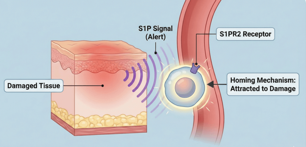
Muse cells do not merely sit in the tissue waiting to be used; they are active repair agents that utilize a specific “homing” mechanism to find damage.
The S1P-S1PR2 Homing Axis
Muse cells have an internal navigation system. When skin is injured, the damaged tissue releases alert signals, such as the lipid signaling molecule sphingosine-1-phosphate (S1P).
Muse cells possess the receptor for this signal (S1PR2). When they are present in the bloodstream or nearby tissue, they “sense” the S1P alert coming from a skin lesion. They travel to that damaged site and adhere to it for swift skin revitalization.
Spontaneous Differentiation
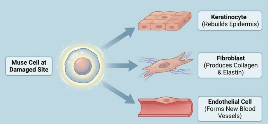
Once they arrive at the site of skin damage, Muse cells get to work. Because they can differentiate into multiple cell types, they can assess what is missing and fulfill that role. In clinical observations of deep wounds, researchers have observed Muse cells transforming into:
- Keratinocytes, which rebuild the epidermis and close the wound surface.
- Fibroblasts, the structural engineers of the skin, are responsible for laying down new collagen and elastin matrices.
- Endothelial cells that form new blood vessels (angiogenesis) are critical for bringing oxygen and nutrients back to the damaged skin.
Clinical Applications for Muse Cells in Dermatology
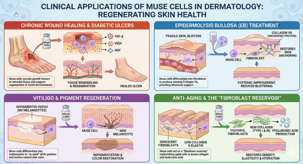
The regenerative capabilities of Muse cells are currently being investigated for several severe dermatological conditions as well as aesthetic concerns.
Chronic Wound Healing and Diabetic Ulcers
One of the most robust areas of Muse cell research is the treatment of diabetic foot ulcers and intractable wounds. In diabetic patients, high blood sugar impairs blood vessel function and immune response, leading to wounds that refuse to heal. This is a leading cause of amputation globally.
Research has demonstrated that when Muse cells are introduced to these environments, they do more than just survive; they remodel the tissue.
A study by Kuroda et al. involved injecting Muse cells into diabetic ulcers, with the data showing the cells reached the dermis and epidermis. The study showed Muse cells secrete high levels of growth factors such as TGF-β (Transforming Growth Factor-beta), VEGF (Vascular Endothelial Growth Factor), and HGF (Hepatocyte Growth Factor). Each of these signaling elements supports skin regeneration.
Epidermolysis Bullosa (EB)
Epidermolysis Bullosa is a genetic condition where the skin is incredibly fragile, blistering at the slightest touch due to a lack of anchoring proteins, including Collagen VII.
Muse cells offer a promising therapeutic avenue because they can differentiate into fibroblasts that naturally produce the missing Collagen VII. Unlike gene therapy, which attempts to edit the patient’s DNA, Muse cell therapy introduces healthy donor cells that naturally produce the glue the skin needs. Preliminary research found that intravenous administration of Muse cells can lead to systemic improvements, as the cells repair blisters.
Vitiligo and Pigment Regeneration
Vitiligo is an autoimmune condition where melanocytes (the cells that give skin its color) are destroyed, leaving white patches. Current treatments involve immune suppression or UV light, which can help manage the symptoms but don’t address the root cause of cell loss.
Because Muse cells can differentiate into ectodermal lineages, they have the capacity to become melanocytes. Studies utilizing 3D skin models have shown that Muse cells can integrate into the basal layer of the epidermis and differentiate into pigment-producing cells. This suggests Muse cell transplantation could potentially “re-seed” the white patches in vitiligo patients with healthy melanocytes, offering a functional restoration of skin color.
Anti-Aging and the “Fibroblast Reservoir”
As our bodies age, the decline of skin quality is largely due to the loss and senescence (aging) of fibroblasts. Fibroblasts are the factories of the skin, forming collagen, elastin, and hyaluronic acid.
Muse cells act as a fresh “fibroblast reservoir.” When applied to aged or sun-damaged skin, they differentiate into new, youthful fibroblasts through two processes:
- Collagen synthesis supports the formation of new fibroblasts, which produce Type I and Type III collagen, restoring density.
- Hyaluronic acid production, which increases hydration levels from within the dermis.
Muse Cells Vs. Lasers and Microneedling
Treatments like CO2 lasers or microneedling are based on the principle of “controlled injury.” By damaging the skin, they elicit a healing response, leading to the formation of collagen.
- The Limitations of Lasers and Microneedling
This process creates scar-like collagen (fibrosis) and requires significant downtime. It also depends entirely on the body’s remaining ability to heal.
- The Muse Advantage
Muse cells do not require injury to work. They target existing damage or aging sites via chemical signals. They rebuild the tissue architecture physiologically, creating healthy dermal tissue rather than scar tissue.
Muse Cells Vs. Dermal Fillers
Injectable fillers physically occupy space to smooth out wrinkles or add volume.
- The Limitations of Dermal Fillers
Fillers are inert. They sit where they are placed and eventually dissolve. They do not improve the biological quality of the skin surrounding them.
- The Muse Advantage
Muse cells are “living fillers.” While they may not provide the immediate, dramatic volume of a synthetic filler, they integrate into the tissue to produce naturally occurring hyaluronic acid and collagen over time, improving the skin’s actual structure and elasticity from the inside out.
Schedule Your Free Muse Cell Skincare Consultation with Miami’s Leading Muse Cell Doctors
At STEMS Health, we’re redefining the future of dermatological care by moving beyond temporary surface improvements to true cellular regeneration.
Whether addressing deep acne scarring, reversing severe sun damage, or reducing fine lines, our approach utilizes the proven power of Dezawa Muse cells to rebuild tissue architecture from within.
Backed by thousands of successful skin treatments, our clinic offers data-backed stress-enduring muse cell therapy for patients seeking lasting skin restoration and aesthetic health. Schedule a free video consultation with our medical team today.
FAQs on Muse Cell Skin Treatments
Can Muse cells actually fill in deep acne scars or “ice pick” scars?
Potentially, this is one of their most promising skincare applications for Muse cells. Deep acne scars represent a loss of dermal tissue that the body failed to replace. Because Muse cells can differentiate into dermis-forming fibroblasts, they can rebuild the tissue matrix from the bottom of the scar upward. Unlike lasers that smooth the surface, Muse cells add biological volume to the depression.
What is the downtime after a Muse cell skin injection?
The downtime after Muse cell skin injections is minimal, comparable to a standard filler injection. You may experience minor swelling, redness, or bruising at the injection sites for 24 to 48 hours.
Because Muse cells are anti-inflammatory by nature, they tend to calm the injection trauma quicker than synthetic fillers. Most patients return to social activities within 2-3 days.
How long do the results last compared to fillers or Botox?
Botox wears off in 3-4 months, and fillers dissolve in 6-12 months. Muse cell therapy aims for biological permanence.
Once the Muse cells differentiate into new fibroblasts, those cells become a permanent part of your skin structure. While they will eventually age just like the rest of your body, the structural repair they perform does not simply “dissolve” or wear off.
Does Muse cell therapy help with sunspots and UV damage?
Photo-aging (sun damage) creates a chaotic environment in the skin where cells function poorly. Muse cells can repair UV-induced damage by replacing the senescent cells that accumulate after sun exposure. Their ability to differentiate into healthy melanocytes can help normalize irregular pigmentation caused by sun damage.
Are there side effects specific to facial injections of Muse cells?
The safety profile is high, but “over-activity” is a theoretical risk being monitored. In rare cases, the introduction of potent stem cells could temporarily cause the skin to feel “active” or flushed as blood flow increases (angiogenesis).
Unlike fillers, there is no risk of the material migrating or forming lumps (granulomas) because the cells integrate seamlessly into the tissue lattice.
Is the Muse cell skincare procedure painful?
The pain level is generally low and managed with topical numbing cream, similar to microneedling or filler injections. If the Muse cells are administered intravenously for systemic skin rejuvenation (such as in eczema or Epidermolysis Bullosa), the procedure is painless aside from the initial needle stick in the arm.
Sources:
Kuroda, Y., Kitada, M., Wakao, S., et al. (2010). “Unique multipotent cells in adult human mesenchymal cell populations.” Proceedings of the National Academy of Sciences (PNAS), 107(19), 8639–8643.
Kinoshita, K., Kuno, S., Ishigami, T., et al. (2015). “Therapeutic Potential of Adipose-Derived SSEA-3-Positive Muse Cells for Treating Diabetic Skin Ulcers.” Stem Cells Translational Medicine, 4(2), 146–155.
Yamada, Y., et al. (2018). “S1P-S1PR2 Axis Mediates Homing of Muse Cells Into Damaged Heart for Recovery.” Stem Cells, 36(7).
Fujita, Y., et al. “Intravenous allogeneic multilineage‐differentiating stress‐enduring cells in adults with dystrophic epidermolysis bullosa: A phase 1/2 open‐label study.” Journal of the European Academy of Dermatology and Venereology, vol. 35, no. 8, 2021, pp. e528–e531. PubMed Central
Wakao, S., et al. (2011). “Multilineage-differentiating stress-enduring (Muse) cells are a primary source of induced pluripotent stem cells in human fibroblasts.” Proceedings of the National Academy of Sciences.
